A recently identified network of vessels within the brain, potentially contributing to the clearance of metabolic waste, could significantly alter our comprehension of brain function and neurodegenerative diseases. If future studies confirm this discovery, it may pave the way for novel therapeutic approaches for conditions such as Alzheimer’s disease.
Per Kristian Eide, a researcher at the University of Oslo not involved in the current study, remarked that such a confirmation would represent a substantial advancement. “If it’s true, this is huge,” Eide stated, suggesting it could mark a paradigm shift in understanding not only neurodegenerative diseases but also conditions like stroke, traumatic brain injury, and fundamental brain processes.
The Brain’s Waste Disposal System
The brain naturally eliminates metabolic byproducts through the glymphatic system. This system comprises channels that surround the brain’s blood vessels, ultimately connecting to the broader lymphatic system. While most imaging techniques have located lymphatic vessels in the brain’s outer protective layers, they have largely failed to detect them within the brain tissue itself.
Discovery of a Novel Vascular Network
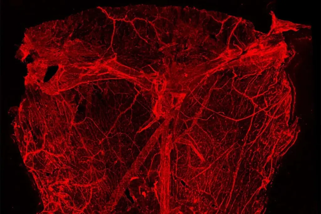
Chongzhao Ran and his colleagues at Harvard University may have identified a previously undiscovered network of lymphatic-like vessels situated inside the brain, with connections to the glymphatic system. Ran described this as his most significant discovery in three decades, calling it a scientist’s dream.
Team member Shiju Gu, also from Harvard, initially observed these structures incidentally. They appeared while Gu was investigating the protein beta-amyloid in brain slices from mice exhibiting Alzheimer’s disease symptoms. Beta-amyloid supports neuronal function but can also form toxic aggregations, a characteristic feature of Alzheimer’s, which might arise from impaired brain drainage.
Subsequent experiments replicated across mice with and without Alzheimer’s symptoms consistently revealed numerous vessel-like structures. These were found in various brain regions, including the cortex, vital for cognitive functions like thinking and problem-solving; the hippocampus, essential for memory formation; and the hypothalamus, which regulates sleep and body temperature.
Ran suggested these structures appear to encircle the brain’s blood vessels and the meningeal lymphatic vessels located in the outer protective membrane. This positioning implies a role in facilitating waste removal through the glymphatic and lymphatic systems.
Human Tissue and Initial Hypotheses
Crucially, the researchers located these tube-like formations in brain samples from an individual who had died with Alzheimer’s disease. Ran also confirmed their presence in brain tissue from someone who did not have the condition.
The research team initially hypothesized that these structures might be a form of lymphatic vessel. They proposed that these vessels could be lined by cells containing or coated with beta-amyloid. Another possibility considered was that the structures represented a form of the protein that could develop into solid fibers, contributing to Alzheimer’s disease but also occasionally found in healthy brains.
Investigating the Nature of the Structures
To clarify their identity, the researchers applied protein markers specific to lymphatic vessels to brain slices from mice. These markers consistently highlighted the tube-like structures, though with less intensity than known lymphatic vessels from the same animals. Based on these findings, the structures were named nanoscale lymphatic-like vessels, or NLVs, and the team concluded they were not a manifestation of beta-amyloid.
However, Eide raised a point of caution regarding the weak staining. He suggested that NLVs might not be lymphatic-like vessels, as such markers can also bind to non-lymphatic tissues. “This is a new kind of structure we’ve not known about before – but it’s unclear, what is this actually?” he questioned.
Christopher Brown from the University of Southampton suggested that these structures could potentially be an artifact generated by the imaging technique itself. He explained that uneven expansion of tissue samples during processing might create the appearance of vessel-like fractures.
Brown indicated that this artifact hypothesis could explain why previous brain imaging studies employing more robust techniques, such as electron microscopy, had not reported the presence of NLVs. Gu mentioned that the team plans to employ electron microscopy in the coming weeks. He also noted that earlier research might have misinterpreted NLVs as axons, which are neuronal projections that share a visual resemblance.
Evidence for Waste Transport and Future Implications
Ran expressed a high degree of confidence in their findings, referencing a separate team study. In this study, fluorescently tagged beta-amyloid within mouse brains appeared to enter adjacent NLVs, suggesting that these structures do indeed transport waste fluid.
If these findings are validated by other research groups, the discovery could significantly advance the understanding of Alzheimer’s disease and other protein-misfolding disorders like Parkinson’s disease. Brown suggested that it could even lead to the development of new treatments, for example, by developing drugs that dilate these vessels to improve waste fluid disposal.