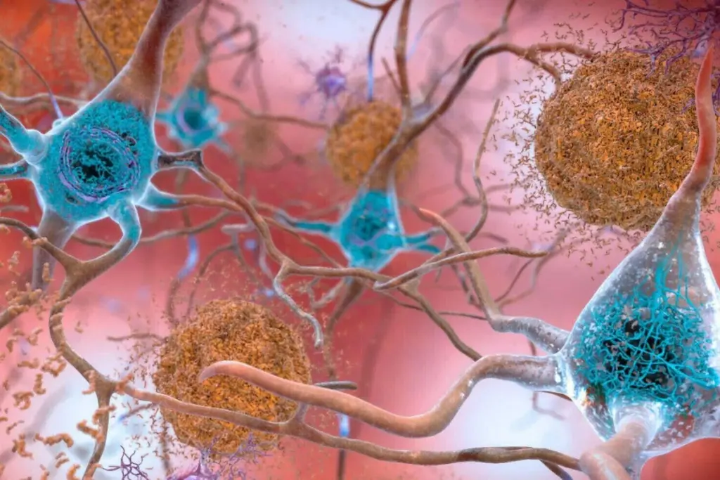
Certain individuals exhibit the physiological changes associated with Alzheimer’s disease, such as the buildup of amyloid plaques and tau tangles in the brain, without ever displaying characteristic symptoms like memory loss. The precise reasons behind this phenomenon, often referred to as resilience, remain a subject of investigation. However, two recent scientific studies are shedding light on potential mechanisms, suggesting that individuals who are resilient possess unique alterations within their brains that may offer protection against cognitive decline.
In Alzheimer’s disease, the accumulation of misfolded proteins, forming amyloid plaques and tau tangles, is broadly understood to be the primary driver of cognitive impairment. Yet, the presence of these pathological hallmarks does not universally manifest as symptomatic disease. Researchers have explored this observed resilience, with a notable 2022 study by Henne Holstege and her team at Amsterdam University Medical Center identifying centenarians who maintained good cognitive function despite the presence of these proteins.
Building on this, Holstege and her colleagues conducted a subsequent study to delve deeper into the mechanisms of Alzheimer’s resilience. Their analysis involved examining brain tissue from 190 deceased individuals. This cohort included 88 individuals diagnosed with Alzheimer’s disease, 53 who showed no signs of the condition at death, and 49 centenarians who did not have Alzheimer’s or any other form of dementia. Among these centenarians, 18 had demonstrated signs of cognitive impairment on assessments performed in the year preceding their death.
Focusing on Key Brain Regions and Protein Accumulation
The researchers specifically investigated the middle temporal gyrus, a brain region identified as one of the earliest sites where amyloid plaques and tau tangles coexist in Alzheimer’s disease. Their findings revealed a subset of 18 centenarians, eight of whom exhibited no cognitive impairment, had amyloid plaque levels comparable to those found in individuals diagnosed with Alzheimer’s. Strikingly, their tau protein levels were similar to those of individuals who had died between the ages of 50 and 99 without the disease.
Holstege suggests that this observation indicates that preventing the accumulation of tau protein is a critical factor in achieving resilience against Alzheimer’s. While amyloid plaques are still linked to cognitive decline, she posits they might create an environment conducive to the spread of tau, thereby triggering Alzheimer’s symptoms. Nevertheless, it is possible for individuals to develop amyloid plaques without significant tau tangle formation. As Holstege notes, “Without amyloid, we don’t see tau spreading.”
Proteomic Analysis Reveals Differential Impact of Tau and Amyloid
Further evidence supporting this distinction emerged from an examination of nearly 3500 proteins within the brains of the study participants. The research indicated that only five of these proteins showed a significant association with the quantity of amyloid plaques. In contrast, a much larger group of approximately 670 proteins were linked to the abundance of tau tangles. Many of these proteins play vital roles in cellular functions such as growth, communication, and metabolism, including the breakdown of waste products. Holstege summarized this finding by stating, “Some things change [in the brain] with amyloid, but everything changes with tau.”
When the researchers focused on tau pathology in the 18 centenarians with elevated amyloid plaques, they discovered that 13 individuals displayed substantial tau spreading, with tangles evident throughout the middle temporal gyrus. Although this pattern of spread mirrors that seen in Alzheimer’s disease, the overall quantity of tau in these individuals remained low.
This distinction is considered significant by researchers. While Alzheimer’s diagnosis incorporates the extent of tau spread throughout the brain, these findings suggest that the accumulation of tau, rather than its mere spread, is the primary driver of cognitive decline. Holstege emphasized this point, stating, “We should really understand that spreading does not necessarily mean abundance.”
Investigating Microglial Function in Resilient Brains
A second study, conducted by Katherine Prater and her colleagues at the University of Washington in Seattle, examined the brains of 33 deceased individuals. This group comprised 10 individuals diagnosed with Alzheimer’s, 10 with no signs of the condition, and 13 classified as resilient. The majority of participants in this cohort were over 80 years old at the time of death, and all had undergone cognitive assessments within a year prior to their passing.
Consistent with the prior study, Prater’s team observed tau spreading, but not significant accumulation, in the brains of individuals with Alzheimer’s resilience. The exact biological pathway for this phenomenon is not yet fully understood, but Prater hypothesizes that microglia, a type of immune cell specialized to the brain, may play a part. Microglia are instrumental in regulating inflammation—a process that is prevalent in Alzheimer’s—as well as maintaining neurons and clearing cellular debris, including amyloid plaques and tau tangles.
Previous research has indicated that microglia can become dysfunctional in Alzheimer’s disease, potentially contributing to neurodegeneration. While the researchers in this study could not directly analyze microglia due to their relative rarity compared to other brain cells, Holstege acknowledged their likely involvement. “But clearly they are involved,” she stated.
Genetic Profiling of Microglia in Resilient Individuals
Prater and her team also performed genetic analysis on the microglia of their cohort, specifically within the dorsolateral prefrontal cortex. This brain region is crucial for high-level cognitive functions such as planning, decision-making, and problem-solving, and it is known to shrink and deteriorate in Alzheimer’s disease.
The analysis revealed that microglia from the resilient individuals exhibited heightened activity in genes associated with messenger RNA transport. Messenger RNA carries genetic instructions for protein synthesis, suggesting that these resilient cells were actively conveying these instructions to protein-production sites. This gene activity in resilient individuals was comparable to that observed in people without Alzheimer’s disease, pointing to a potential disruption in this process as a characteristic of the condition. “If that process gets interrupted, we know that is really bad for cells,” Prater commented.
Furthermore, microglia from resilient individuals showed decreased activity in genes involved in energy metabolism when compared to those from patients with Alzheimer’s. This metabolic activity pattern in resilient individuals resembled that of people without the disease, implying that microglia in Alzheimer’s patients may expend more energy, potentially due to increased inflammatory responses, as Prater suggested. This finding aligns with the understanding that brain inflammation can impair neuronal connections and lead to cell death.
Potential for New Therapeutic Strategies
“Both these studies suggest that the human brain possesses mechanisms for mitigating tau burden,” Prater concluded. A deeper understanding of these natural protective processes could pave the way for novel therapeutic interventions aimed at preventing Alzheimer’s disease, rather than solely managing its progression. “We are certainly not close to a therapeutic yet, but I think that biology is showing us there is hope [and] there is promise,” she added.